
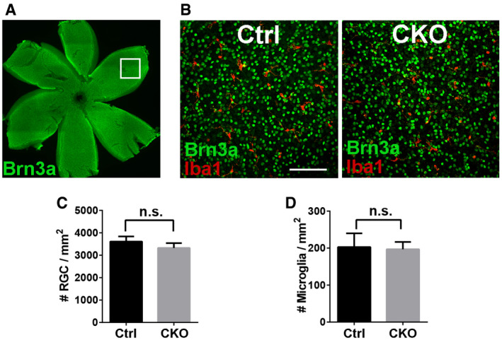
Figure EV4. Deleting microglial Gpr56 has no significant effect on RGC density in P5 retina.
-
AWhole‐mount retinal staining of RGC using Brn3a antibody. The box indicates an example of imaging area in the peripheral retina.
-
BRepresentative images of RGC and microglia staining using Brn3a and Iba1 antibodies in retina. Scale bar, 100 μm.
-
CQuantification of RGC density in controls and CKO. N = 3, P = 0.187.
-
DQuantification of retinal microglia density in controls and CKO. N = 3, P = 0.810.