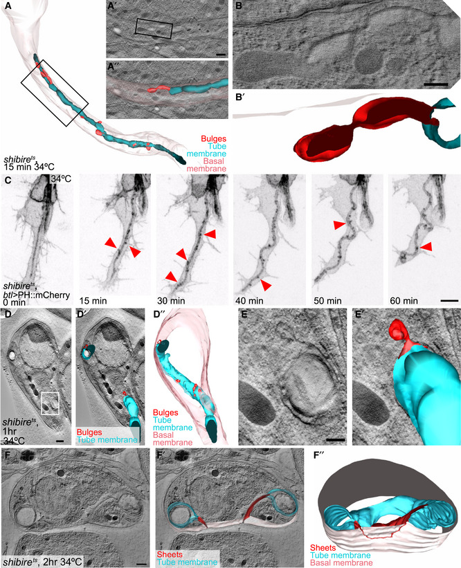
Figure EV3. Effects of dynamin inactivation on membrane morphology.

-
A, BOlder shibire ts terminal cell that had already formed a long branch and tube before dynamin inactivation, fixed 15 min after inactivation. (A) Reconstruction of the entire cell. (A′, A″, B, B′) Higher magnification details of the cell, tomograms and reconstructions.
-
Cshibire ts terminal cell expressing PH::mCherry. Red arrowheads point to puncta of fluorescent material at the tube membrane.
-
D–FTEM tomograms and 3D reconstructions of older shibire ts terminal cells similar to (A), but after 1 h (D–E) and 2 h (F) at restrictive temperature. Box in (D) is magnified in (E). (F″) The position at which the sheet between apical and basal is connected to the basal membrane is traced in red on the outside view of the basal membrane. The cells shown in (D–F) were found and acquired without the CLEM approach.
