
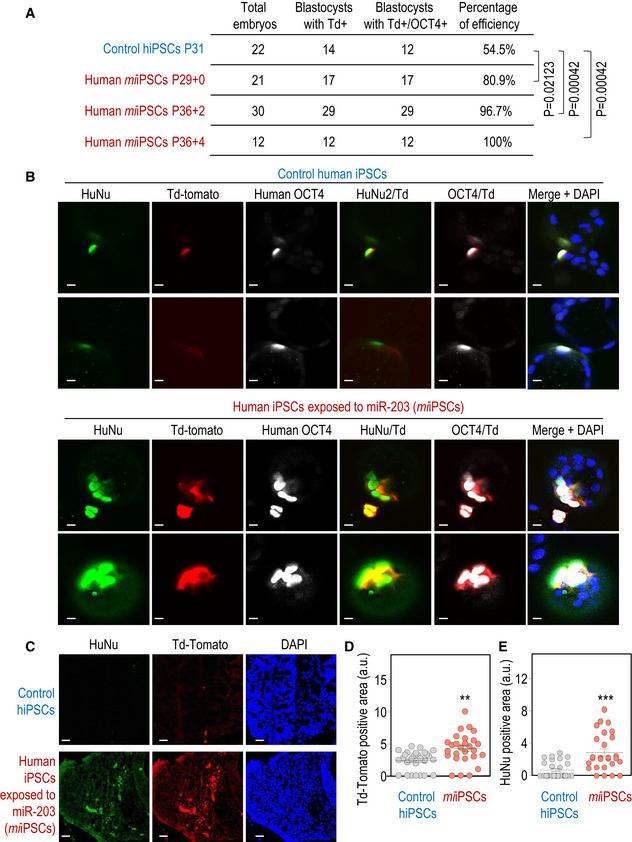
Figure 3. miR‐203‐exposed human iPSCs efficiently contribute to human–mouse interspecies chimeras.
-
ASummary of chimera assays in which one single Td‐Tomato fluorescent‐labeled human iPSC, either control or transiently transfected with miR‐203‐expressing vectors (miiPSCs), was injected into 8C‐stage mouse embryos and their contribution analyzed 48‐60 h later. P‐values are indicated in the figure (Student's t‐test).
-
BRepresentative images showing the contribution of human control iPSCs or miiPSCs to mouse blastocysts. These structures were co‐immunostained with (human) anti‐OCT4 and anti‐HuNu antibodies. Td‐Tomato, red; Scale bars, 50 μm.
-
CInterspecies chimera assays in which the 8C‐stage mouse embryos indicated in (A, B), injected with one single human iPSCs and cultured in vitro for 48 h to reach the blastocyst stage, were then transfected to 2.5 days post‐coitum pseudo‐pregnant females. The post‐implantation mouse conceptuses were dissected at the E9.5 developmental stage and analyzed by immunofluorescence. Representative images showing the integration of control human iPSCs (top) or miiPSCs (bottom) into mouse E9.5 embryos. Anti‐human HuNu antibody was co‐stained with Td‐Tomato direct fluorescence to detect human cells in E9.5 mouse embryos. Scale bars, 20 μm.
-
D, EQuantification of the Td‐Tomato‐positive (D) or HuNu‐positive (E) area in 40‐50 different cryosections of these embryos randomly and blindly selected for IF analysis. Data are mean ± SEM **P < 0.01; ***P < 0.001 (Student's t‐test).
Source data are available online for this figure.