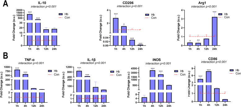
Fig. 2.
Effects of Hb on the polarization of primary microglia. Microglia were cultured in medium containing 20 μM Hb for 1–24 h. a The mRNA expression of anti-inflammatory markers (IL-10, CD206, and Arg1) (n = 3). b The mRNA expression of pro-inflammatory markers (TNF-α, IL-1β, iNOS, and CD86) (n = 3). The data are shown as the relative changes of the experimental group versus the control group (baseline) and were analyzed by two-way ANOVA followed by Sidak post hoc multiple comparison, *p < 0.05, **p < 0.01, and ***p < 0.001. n is the number of independent cell samples

