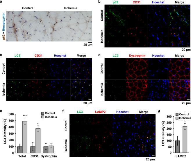
Fig. 3. Autophagy was induced in endothelial cells of HLI mice.
a Immunostaining of p62 was performed using quadriceps tissue from HLI mice. Nuclei were counterstained with hematoxylin. Scale bar = 25 μm. b–d, f Double immunofluorescence staining was performed using specific antibodies as indicated. p62 and CD31 (b); LC3 and CD31 (c); LC3 and dystrophin (d); and LC3 and LAMP2 (f). Nuclei were stained with Hoechst 33342. Scale bar = 20 μm. e, g The total LC3 signal and the overlapped LC3 signal with the indicated protein were quantified using Image J software and plotted. *p < 0.05, ***p < 0.001 versus control.

