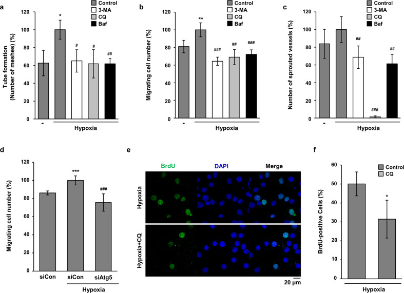
Fig. 5. Autophagy inhibitors decreased the angiogenic activities of endothelial cells under hypoxia.
a HMEC-1 cells were incubated on Matrigel under hypoxia with 3-MA (2 mM), CQ (25 μM), or Baf-A1 (10 nM) for 24 h. The tube formation assay was quantified by counting the meshes from three independent experiments using ImageJ software. *p < 0.05 versus normoxia; #p < 0.05 and ##p < 0.01 versus the hypoxia vehicle. b To compare the motility of HMEC-1 cells, a wound migration assay was performed under hypoxia with autophagy inhibitors as indicated. The number of migrated cells from the reference line was counted, quantified, and plotted. **p < 0.01 versus normoxia; ##p < 0.01 and ###p < 0.001 versus the hypoxia vehicle. c Rat aortic rings were incubated on Matrigel under hypoxia with autophagy inhibitors as indicated. Sprouted microvessels from the aorta were quantified and plotted. ##p < 0.01 and ###p < 0.001 versus the hypoxia vehicle. d HMEC-1 cells were transfected with siControl (siCon) or siAtg5, and then a wound migration assay was performed under hypoxia. e The conditioned media were collected from hypoxia and CQ-treated HMEC-1 cells. C2C12 cells were treated with the conditioned media for 24 h. Proliferative properties of C2C12 cells were evaluated by BrdU incorporation assay and nuclei were stained with Hoechst 33342. Scale bar = 20 μm. f The data are expressed as means ± S.D. for three determinations in three independent experiments. *p < 0.05.

