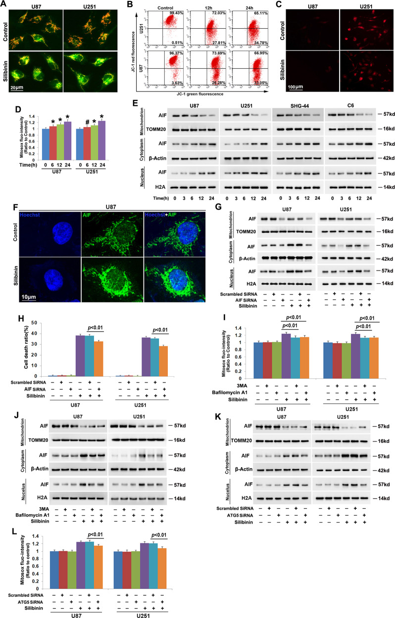
Fig. 2. Autophagy contributed to silibinin-induced mitochondria damage.
a Representative images of the cells stained with JC-1 under fluorescence microscope showed that silibinin treatment resulted in obvious reduction of red fluorescence in U87 and U251 cells. b Flow cytometry analysis combined with JC-1 staining confirmed that silibinin induced time-dependent dissipation of mitochondria membrane potentials. c Representative images of the cells stained with Mitosox red under fluorescence microscope showed that the red fluorescence exhibited by Mitosox red was apparently stronger in silibinin-treated cells than that in control group. d Statistical analysis of the red fluorescence intensity exhibited by Mitosox red proved that silibinin triggered mitochondrial accumulation of superoxide in a time-dependent manner. e Western blotting analysis revealed that silibinin induced AIF translocation from mitochondria to nuclei in a time-dependent manner. f Representative images acquired by confocal microscopy combined with immunochemical staining showed that silibinin induced accumulation of AIF in the nucleus of U87 cell. g Silibinin-induced accumulation of AIF in nuclear fraction was decreased in the cells transfected with AIF SiRNA. h LDH release assay proved that knockdown of AIF prevented silibinin-induced glioma cell death. i Statistical analysis of the red fluorescence intensity exhibited by Mitosox red showed that silibinin-induced mitochondrial accumulation of superoxide was significantly inhibited in the presence of 3MA or bafilomycin A1. j Western blotting proved that knockdown of ATG5 with SiRNA prevented silibinin-induced AIF translocation from mitochondria to nuclei. k Western blotting revealed that silibinin-induced nuclear translocation of AIF was suppressed when ATG5 was knocked down with SiRNA. l Knockdown of ATG5 with SiRNA abrogated the improvement of mitochondrial superoxide induced by silibinin.

