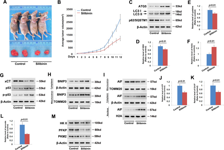
Fig. 7. Silibinin inhibited glioma cell growth in vivo.
a Representative images of the mice with xenografted gliomas and the removed tumors. b Statistical analysis of silibinin-induced changes in tumor volumes. The average tumor size was 2750 ± 406 mm3 in control group, which reduced to 1225 ± 157 mm3 after being treated with silibinin for 12 days (n = 5). c Western blotting analysis revealed that silibinin upregulated ATG5 and LC3-II, but downregulated p62. d–f Silibinin treatment resulted in depletion of GSH and cysteine, but improvement in H2O2. g Western blotting analysis showed that silibinin downregulated xCT, but upregulated p53 and phospho-p53. h Silibinin triggered BNIP3 upregulation and accumulation on mitochondria. i Silibinin promoted AIF translocation from mitochondria to nuclei. j–l Silibinin significantly induced depletion of ATP, glucose-6-phosph, and pyruvate. m Silibinin treatment resulted in downregulation of HK II, PFKP, and PKM2. The values are expressed as mean ± SEM (n = 5 per group).

