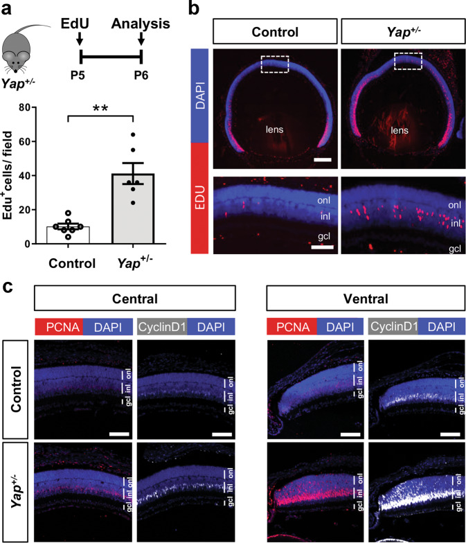
Fig. 2. Prolonged proliferation of retinal progenitors at postnatal stages in Yap+/− mice.
a Timeline diagram of the experimental procedure used in b. Wild-type (Control) or Yap+/− mice were injected with EdU at P5 and analyzed 24 h later. b P6 retinal sections labelled for EdU (red) and stained with DAPI (blue). The delineated areas are enlarged in the bottom panels. Scatter plot with bars represents the number of EdU+ cells per field (250 × 250 µm). Means ± SEM from seven control retinas and six Yap+/− retinas are shown. c P6 retinal sections immunostained for PCNA (red) or Cyclin D1 (grey). Both central and peripheral ventral regions are shown. Nuclei are DAPI counterstained (blue). inl: inner nuclear layer, onl: outer nuclear layer, gcl: ganglion cell layer. Statistics: Mann–Whitney test, **p ≤ 0.01. Scale bar: 200 µm (b) and 50 µm (c and enlarged panels in b).

