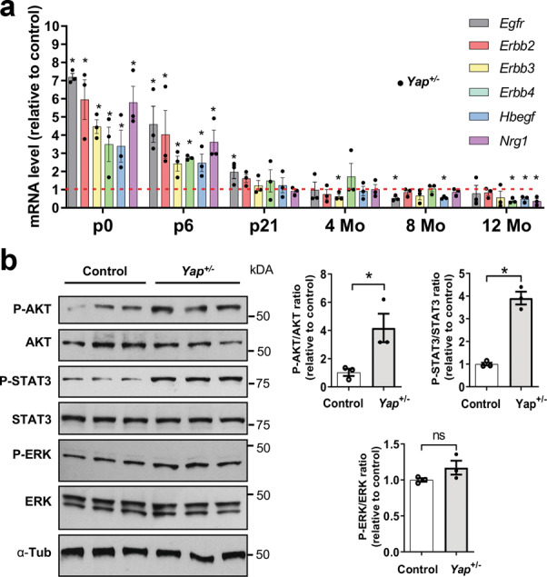
Fig. 3. EGFR pathway activation in the retina of Yap+/− postnatal mice.

a RT-qPCR analysis of various EGFR pathway signaling gene expression (Egfr, Erbb2, Erbb3, Erbb4, Hbegf, and Nrg1), relative to wild-type controls (dashed lines) (n = 3 biological replicates per condition). b Analysis of protein expression levels of EGFR signaling pathway components at P6 by western blot. Quantifications of p-AKT/AKT, p-STAT3/STAT3, and p-ERK/ERK ratios are relative to controls (n = 3 biological replicates per condition). All values are expressed as the mean ± SEM. Statistics: Mann–Whitney test, *p ≤ 0.05, ns nonsignificant.
