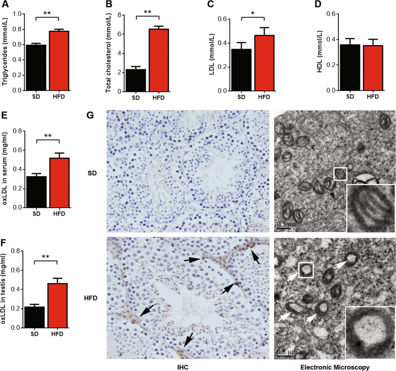
Fig. 2. oxLDL level is significantly elevated in the serum and testes of HFD mice.
Serum triglyceride (a), serum total cholesterol (b), serum LDL (c), and serum HDL (d) levels in mice were detected by a biochemical analyzer. Serum oxLDL (e) and testicular oxLDL (f) levels were measured by ELISA. *P < 0.05, **P <0.01, compared with SD-fed mice. (g) Immunohistochemical staining indicated that oxLDL accumulated in the testicles in SD- and HFD-fed mice, and the arrows show sites of positive expression (n = 8 per group; scale bar, 20 μm). Transmission electron microscopy showing the ultrastructural characteristics of mitochondria of Leydig cells in SD- and HFD-fed mice. The arrows show the absence of mitochondrial cristae (n = 8 per group; scale bar, 0.5 μm).

