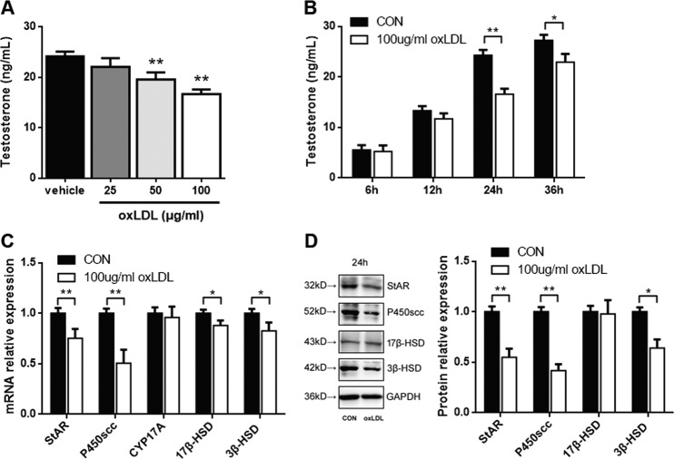
Fig. 3. oxLDL inhibits testosterone production in primary Leydig cells.
Cells were treated with various concentrations of oxLDL for 24 h (a) or with 100 μg/mL oxLDL for different time points (b). Then, the culture medium was collected and assayed for testosterone production. c Cells were treated with 100 μg/mL oxLDL for 24 h, and the mRNA expression of StAR, P450scc, CYP17A, 17β-HSD, and 3β-HSD was examined by qRT-PCR. d The protein expression of StAR, P450scc, 17β-HSD, and 3β-HSD was detected by western blotting. Quantitation of testosterone synthesis-related proteins and enzyme expression was determined by normalization to the internal control GAPDH. The vehicle with no oxLDL treatment was used as a control. The data are presented as the mean ± standard deviation of at least three independent experiments. *P < 0.05, **P < 0.01, compared with the control.

