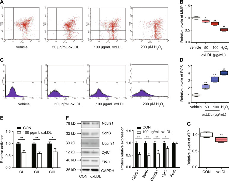
Fig. 4. oxLDL inhibits mitochondrial function in primary Leydig cells.
Cells were treated with various concentrations of oxLDL or 200 μM H2O2 for 24 h, and then the mitochondrial membrane potential (MMP) (a, b) and intracellular ROS (c, d) were assayed by flow cytometry. H2O2 as a positive control. e Activities of mitochondrial complexes I, II, and III (CI, CII, and CIII) were assessed by spectrophotometry. f The expression of respiratory chain proteins, including Ndufs1 (a subunit of CI), SdhB (a subunit of CII), Uqcrfs1 (a subunit of CIII), Fech (a matrix enzyme ferrochelatase), and CytC (an intermembrane space protein cytochrome C), was detected by western blotting. Quantitation of mitochondrial proteins expression was determined by normalization to the internal control GAPDH. g The level of Intracellular ATP were detected. The vehicle with no oxLDL treatment was used as a control. The data are presented as the mean ± standard deviation of at least three independent experiments. *P < 0.05, **P < 0.01, compared with the control.

