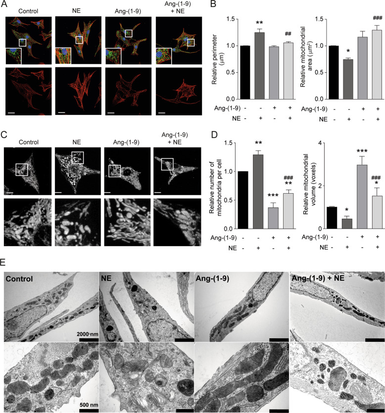
Fig. 2. Angiotensin-(1–9) prevents mitochondrial fission elicited by norepinephrine.
a Representative confocal images of cardiomyocytes treated with 100 μM angiotensin-(1–9) for 6 h and then stimulated with 10 μM of norepinephrine (NE) for 24 h. Cardiomyocytes were stained with rhodamine phalloidin to detect sarcomeric structures and immunolabeled for the mtHsp70 protein to identify the mitochondrial network. Scale bar: 25 μm. b Quantitative analysis (n = 4) of relative cellular perimeter and mitochondrial area of cardiomyocytes. c Mitochondrial morphology of cardiomyocytes pretreated as in a. Scale bar: 2 μm. Bottom panels represent a ×13.5 magnification. d Relative number of mitochondria per cell (left) and mean mitochondrial volume (right) were quantified as mitochondrial dynamics parameters (n = 4). *p < 0.05; **p < 0.01 and ***p < 0.001 vs. control; ##p < 0.01 and ###p < 0.001 vs. NE. e Representative transmission electron microscopy images from angiotensin-(1–9)- and NE-treated cells showing two different magnifications of the same cells.

