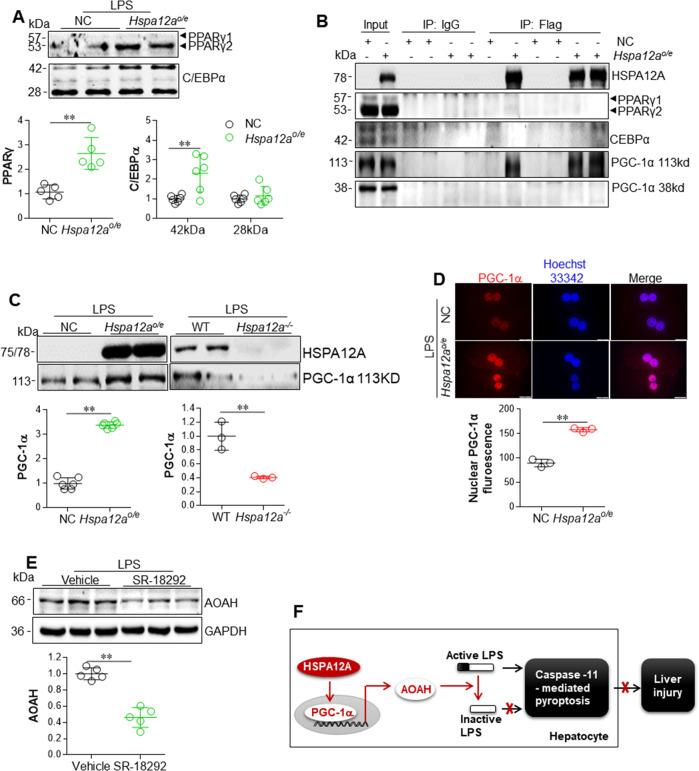
Fig. 8. HSPA12A upregulated AOAH expression through interaction with PGC-1α in hepatocytes.
a HSPA12A increased nuclear contents of PPARγ and C/EBPα in LPS-treated hepatocytes. WT primary hepatocytes were infected with Hspa12a-adenovitus to overexpress HSPA12A (Hspa12ao/e). WT hepatocytes infected with empty virus served as negative controls (NC). After incubation with LPS for 6 h, the nuclear fractions were prepared for immunoblotting with the indicated antibodies. Data are mean ± SD, **P < 0.01 by Student’s two-tailed unpaired t test. n = 5 for PPARγ group, n = 6 for C/EBPα group. b Interaction between HSPA12A and PGC-1α in hepatocytes. Primary WT hepatocytes that overexpressing the flag-tagged HSPA12A (Hspa12ao/e) were incubated with LPS for 6 h. Primary hepatocytes infected empty virus served as negative controls (NC). Cellular protein extracts were immunoprecipitated with primary antibody for flag. The immunoprecipitates were blotted with the indicated antibodies. Protein extracts without immunoprecipitation (input) served as positive controls, and immunoprecipitates from IgG incubation served as negative controls. Note that only PGC-1α was recovered in flag-tagged HSPA12A immunoprecipitates. c Nuclear PGC-1α content was increased by HSPA12A overexpression but decreased by HSPA12A deficiency. Hspa12ao/e primary hepatocytes and its NC controls, Hspa12a−/− primary hepatocytes and its WT controls were treated with LPS for 6 h. Nuclear fractions were prepared for immunoblotting against PGC-1α and HSPA12A. Data are mean ± SD, **P < 0.01 by Student’s two-tailed unpaired t test. n = 6/group for left panels and n = 3/group for right panels. d HSPA12A increased nuclear PGC-1α abundance in LPS-treated hepatocytes. Hspa12ao/e and NC primary hepatocytes were treated with LPS for 6 h. PGC-1α in nuclei was examined by immunostaining. Hoechst 33342 was used to counter staining nuclei. Data were expressed as fluorescence intensity/nucleus. Scale bar = 20 μm. Data are mean ± SD, **P < 0.01 by Student’s two-tailed unpaired t test. n = 3/group. e Inhibition of PGC-1α decreased AOAH expression in Hspa12ao/e hepatocytes. Hspa12ao/e primary hepatocytes were treated with PGC-1α inhibitor SR-18292 or vehicle for 12 h followed by incubation with LPS for 6 h. AOAH expression was examined by immunoblotting. Data are mean ± SD, **P < 0.01 by Student’s two-tailed unpaired t test. n = 5/group. f Mechanism scheme. By directly binding to PGC-1α, HSPA12A promotes PGC-1α translocation to nuclei, thereby promotes AOAH expression for LPS inactivation, and ultimately leads to hepatic protection through inhibition of Caspaase11-mediated pyroptosis of hepatocyte.

