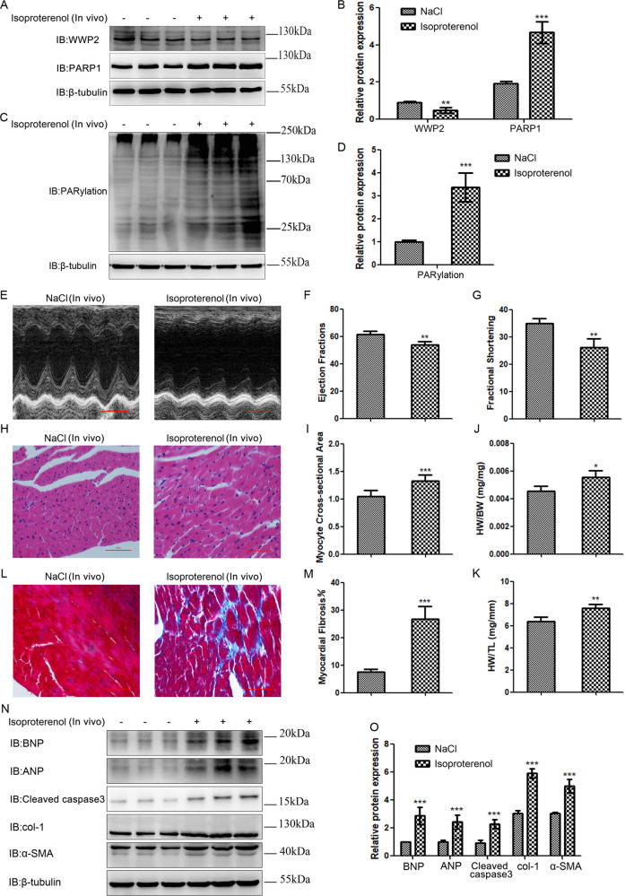
Fig. 4. WWP2 level significantly decreased, while PARP1 and PARylation levels markedly increased in ISO-induced cardiac remodeling in mice.
a, c In C57BL/6 mice following 2 weeks of ISO or NaCl infusion in vivo, western blotting was carried out to assess the expression of WWP2 and PARP1, and PARylation. b, d Data are shown as mean ± SD (each group of mice, n = 6; **P < 0.01, ***P < 0.001, unpaired Student’s t test). e Echocardiography (scale bar: 2 mm), (f) ejection fraction, and (g) fractional shortening were used to assess heart failure. h H&E staining (scale bar: 50 µm), (i) relative myocyte cross-sectional area, (j) heart weight to body weight ratio, and (k) heart weight to tibia length ratio were used to assess myocardial hypertrophy. l Masson trichrome staining (scale bar: 50 µm) and (m) fibrotic area were used to assess myocardial fibrosis. Data (e–m) are shown as mean ± SD (each group of mice, n = 6; *P < 0.05, **P < 0.01, ***P < 0.001, unpaired Student’s t test). n Western blotting was carried out to assess the expression of markers of myocardial hypertrophy, heart failure, and myocardial fibrosis (BNP, ANP, cleaved caspase3, col-1, and α-SMA). o Data are shown as mean ± SD (each group of mice, n = 6; ***P < 0.001, unpaired Student’s t test).

