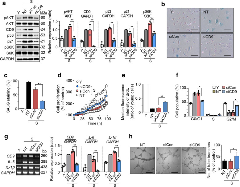
Fig. 1. Knockdown of CD9 in senescent cells rescues cellular senescence.
Senescent HUVECs (PD > 50) were transfected with CD9 or negative control siRNA and then incubated for 6 days at 37 °C. a The levels of pAKT, AKT, CD9, p53, p21, pS6K, and S6K proteins by western blotting and their relative levels. b SAβG staining (blue). Scale bar: 20 μm. c The percentages of SAβG positive cells. d Cell proliferation measured by live-cell time-lapse microscopy and cell counting. e BrdU incorporation measured by flow cytometry. f Cell cycle analysis measured by flow cytometry. g The expression levels of IL-6 and IL-1β mRNAs by RT-PCR and their relative levels. h Tube formation in HUVECs. Scale bar: 200 μm. Representative data are shown and the values are the means ± SD of three independent experiments. Y young cells, S senescent cells, NT not treated, siCon negative control siRNA, siCD9 CD9 siRNA; *p < 0.05 and **p < 0.01.

