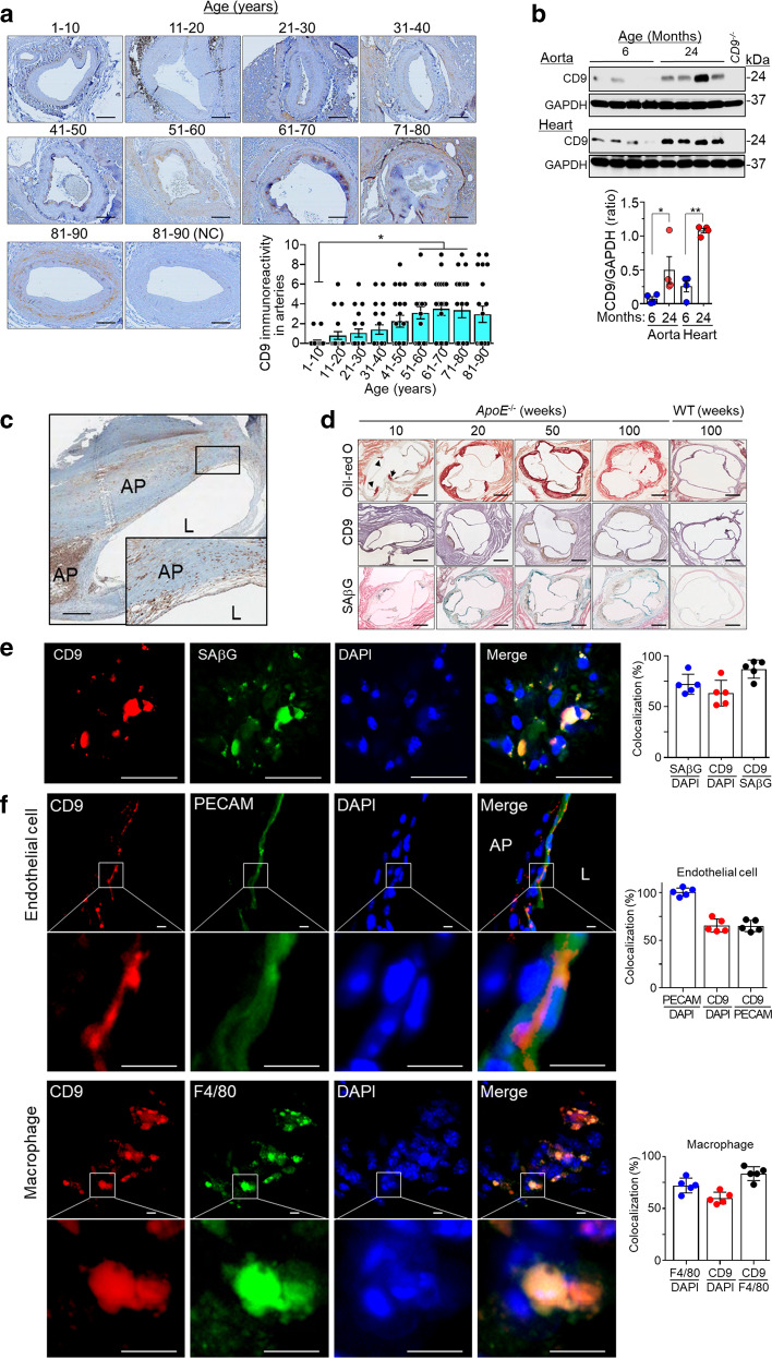
Fig. 4. Upregulation of CD9 in human and rat arterial tissues with age and in atherosclerotic lesions of human carotid arteries and aortic sinuses in ApoE−/− mice.
a CD9 immunostaining (brown) and CD9 immunoreactivity in human arterial tissues with age in each group (each, n = 20). Scale bar: 200 μm. b Levels of CD9 protein measured by western blotting in the aortas and the hearts of 6- and 24-months old rats. c Representative CD9 immunostaining (brown) in atherosclerotic lesions in human carotid arteries (n = 6). Scale bar: 500 μm. d Staining for Oil-red O (red), CD9 immunoreactivity (brown), and SAβG (blue) in aortic sinus sections from ApoE−/− mice with age (each, n = 5). Scale bar: 200 μm. e Immunofluorescence staining and colocalization of CD9 with SPiDER-βGal, a fluorescence marker of SAβG in frozen tissue sections of atherosclerotic lesions of ApoE−/− mice (n = 5). Scale bar: 30 μm. f Immunofluorescence staining and colocalization of CD9 with PECAM, an endothelial cell marker, and F4/80, a macrophage marker in frozen tissue sections of atherosclerotic lesions of ApoE−/− mice (n = 5). Scale bar: 5 μm. CD9−/− CD9 deficient mice, L lumen of artery, AP atherosclerotic plaque, ApoE−/− apolipoprotein E deficient mice, WT C57BL/6 mice, NC without CD9 primary antibody; *p < 0.05 and **p < 0.01.

