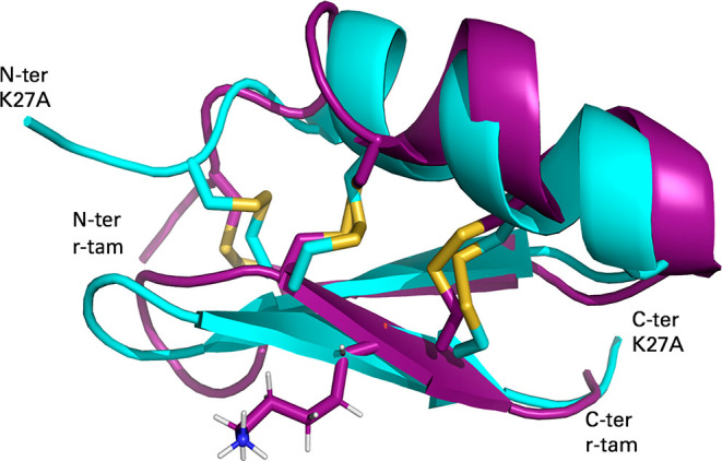
Figure 2.

3D NMR structure of r-tam and K27A mutant. Alignment of mutant (cyan, PDB entry 6D9P) and r-tam (purple, PDB entry 2LU9) from amino acids C3-V29; the K27 amino acid is shown in sticks. K27A is the mutant with the greatest differences in HN protons compared to r-tam. Despite the difference in chemical shifts, both structures conserve the topology.
