Fig. 4.
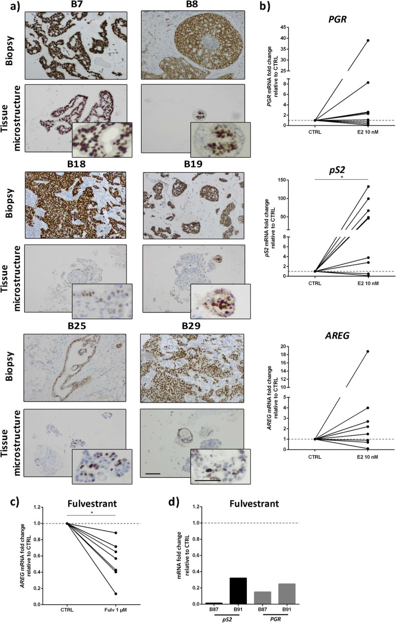
Estrogen Receptor α (ER) expression and functionality were maintained in alginate encapsulated tissue microstructures up to 1 month of culture. a Immunohistochemistry detection of ER in biopsy (top row) and encapsulated tissue microstructures culture for a month (bottom row) (scale bars: 200 μm for low magnification and 100 μm for high magnification). b Encapsulated tissue microstructures were cultured for 3 days in depleted medium and stimulated with 17-β-estradiol; expression of ER downstream target genes was assessed by RT-qPCR (amphiregulin - AREG, progesterone receptor - PGR and protein PS2 - pS2, N = 9). Data are shown as fold change in gene expression upon 17-β-estradiol challenge relatively to vehicle-exposed control (CTRL). c, d Encapsulated tissue microstructures were cultured for 3–5 days in complete medium, before challenge with fulvestrant for 2 weeks; ER downstream targets were assessed by RT-qPCR (AREG, PGR and pS2, N = 7). Data are shown as fold change in gene expression upon fulvestrant challenge relatively to vehicle-exposed control. Statistical analysis was performed by the Mann-Whitney test (*p-value < 0.001)

