
Figure 1.
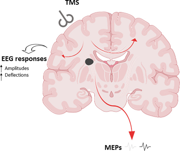
TMS and post-stroke injury: coronal image at the level of the basal ganglia. MEPs may be absent or weak after TMS but EEG responses reveal altered cortical activity post-stroke. When the patient does not present with MEPs, a combination of TMS-evoked EEG allows monitoring of cortical activity evoked by ipsilesional TMS. This can provide accurate prognosis and identify pathways for personalized interventions. Both high-resolution MRI and TMS–EEG approaches reveal impaired integration in the connectome in both hemispheres but less pronounced in the contralateral hemisphere affected by stroke.