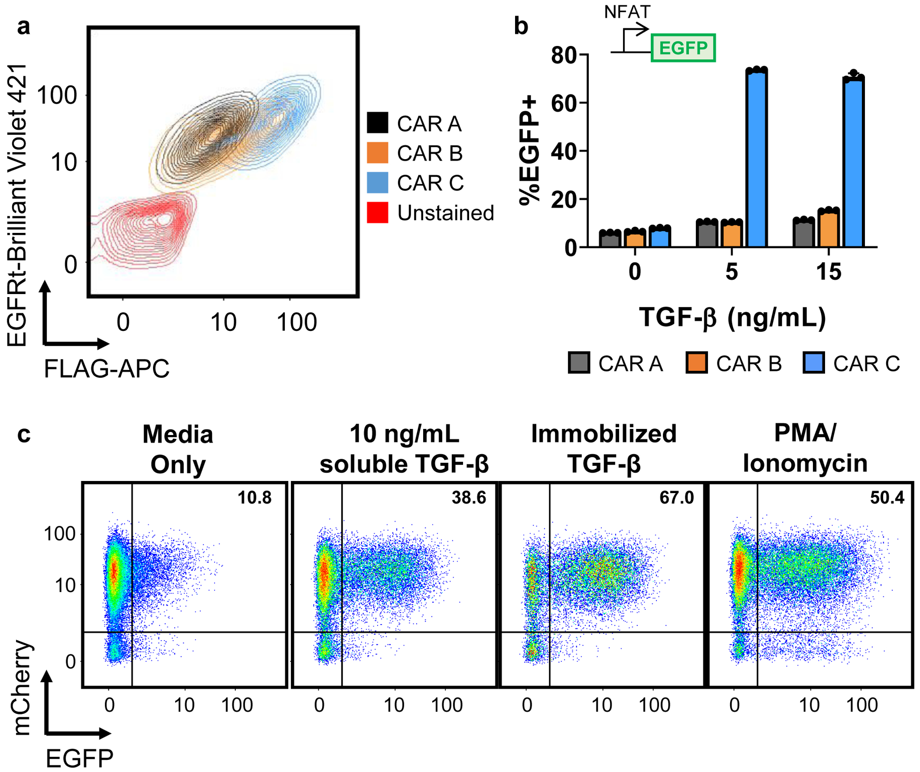
Fig. 2. Troubleshooting CAR expression and effector activity.

a, Different TGF-β scFvs with increasing TGF-β–binding affinities were used to construct CARs A, B, and C, respectively. CARs were fused to an N-terminal FLAG tag, and connected via a T2A “self-cleaving” peptide to truncated EGFR (EGFRt). As such, FLAG staining allows quantification of CAR surface expression while EGFRt staining indicates transduction efficiency. When expressed in Jurkat cells, all three CAR designs transduced well and expressed on the cell surface, albeit with varying surface-localization efficiencies. b, When tested in Jurkat NFAT reporter cell lines, only CAR C produced a clear response at the tested levels of TGF-β concentration. Data points from n = 3 samples are shown with means ± 1 standard deviation. c, NFAT reporter cells transduced to express an mCherry-fused TGF-β CAR were exposed to media alone, 10 ng/mL soluble TGF-β, immobilized TGF-β, or 50 ng/mL PMA and 1 μM ionomycin. TGF-β was immobilized in cell-culture wells by covering the well with 100 ng/mL TGF-β at room temperature for 30 min, washing once with PBS, then allowing the well to dry.
