
Figure 3.
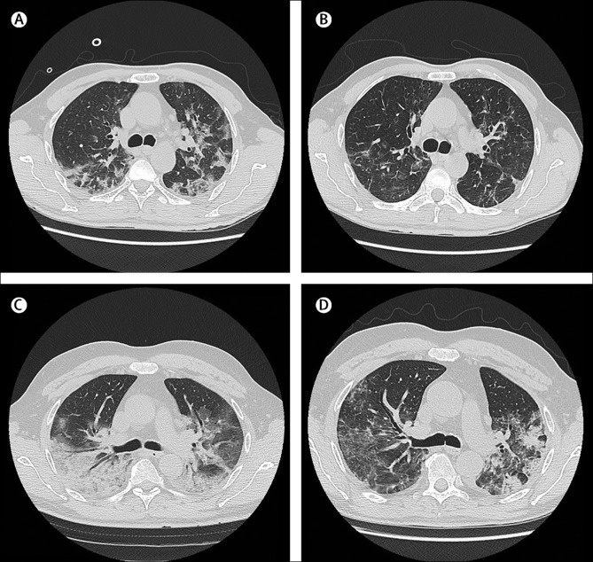
Radiographic findings in two patients in the mavrilimumab group
Lung CT scans of a man aged 58 years at day 0 (A) and at discharge from hospital on day 7 (B). At day 0, the patients was febrile, receiving oxygen through a face mask, with fraction of inspired oxygen (FiO2) of 0·4, partial pressure of oxygen (PaO2) of 86 mm Hg, lactate dehydrogenase (LDH) concentration of 374 U/L, and C-reactive protein (CRP) concentration of 100 mg/L. The day 0 lung CT scan shows presence of bilateral, blurred ground-glass opacities with crazy paving pattern and small dense consolidation areas. The CT scan at discharge (afebrile, on room air, oxygen saturation of 98%, LDH normalised, and CRP concentration of 12·5 mg/L), shows reduction and regression of these findings. Lung CT scans of a man aged 56 years at day 0 (C) and at discharge from hospital on day 14 (D). At day 0, the patient was febrile, receiving high-flow oxygen through a face mask with reservoir bag and continuous positive airway pressure 12 h per day, PaO2 of 176 mm Hg, LDH concentration of 944 U/L, and CRP concentration of 177 mg/L. The day 0 lung CT scan shows extensive involvement of the right lung with a posterior large consolidation area and aerial bronchogram; ground-glass opacities and crazy paving pattern are predominant on the left side. The CT scan at discharge (afebrile, on room air, oxygen saturation of 98%, LDH normalised, and CRP concentration of 28·2 mg/L), shows improvement in lung involvement.