. 2020 Mar 16;42(4):403–419. doi: 10.1590/1516-4446-2019-0741

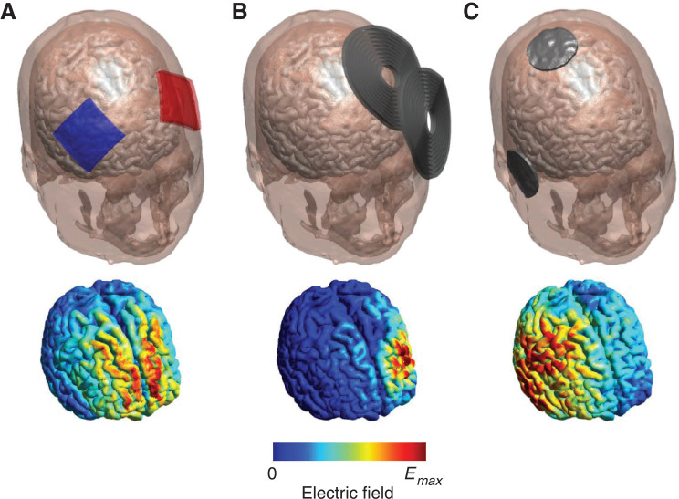
Figure 2. Examples of NIN techniques (top panel) and the corresponding electric field distribution in the brain (bottom panel): A) tDCS using 5 × 5 cm electrodes placed over the bilateral DLPFC; electrodes are colored red and blue to distinguish anode (red) vs cathode (blue). B) TMS using the MagVenture B70 coil over the left DLPFC. C) Right unilateral ECT; conventional ECT uses a bipolar waveform and therefore does not distinguish between anodal vs. cathodal electrodes. Electric field strengths are normalized to their respective maximum value (Emax); absolute field strengths are very different across the modalities (< 1 V/m for tDCS to > 100 V/m for TMS and ECT). Figure produced using SimNIBS software.21 DLPFC = dorsolateral prefrontal cortex; ECT = electroconvulsive therapy; NIN = non-implantable neuromodulation; tDCS = transcranial direct current stimulation; TMS = transcranial magnetic stimulation.

Figure 2