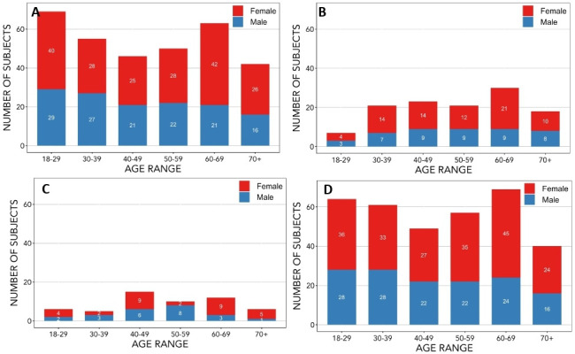
Figure 1.

Distribution of age at study entry and sex of participants recruited up to December 2018. The age and sex distribution of participants has been provided for (A) all baseline MR scans completed, (B) follow-up MR scans completed, (C) participants whose scans were completed with phase 1 MR acquisition protocol and (D) participants whose scans were completed with phase 2 MR acquisition protocol. Phase 1 MR acquisition protocol was used only for baseline scans; phase 2 MR acquisition protocol was used for some baseline and all follow-up scans. The exact number of participants for each sex in a given age category are indicated in the appropriate portion of the bar representing that category. MR, magnetic resonance
