Figure 7.

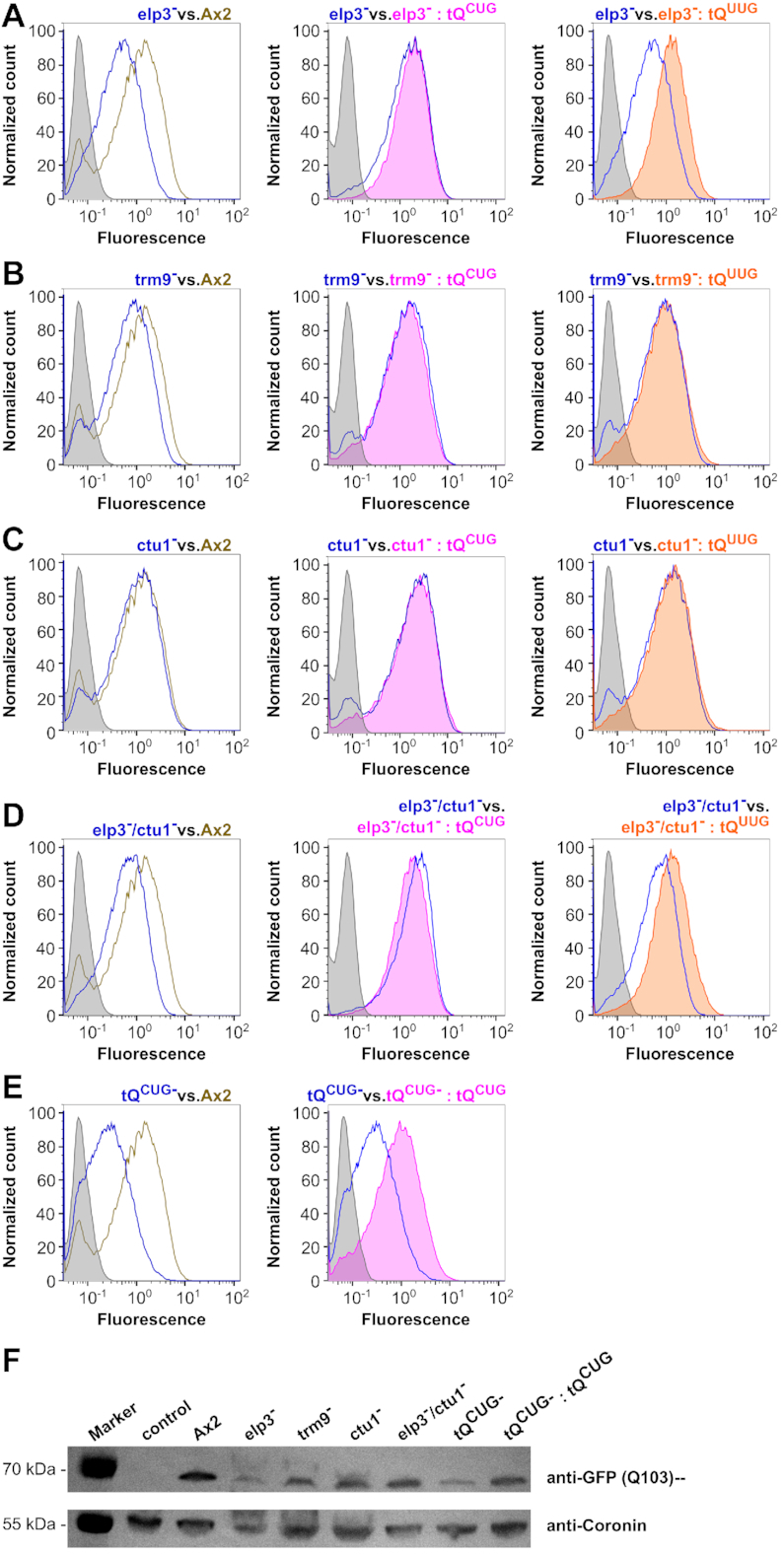
Expression of GFP with the Q103 leader sequence. Representative flow cytometry detected fluorescence profiles of Q103-GFP in Ax2 wild type and elp3− (A), trm9− (B), ctu1− (C), elp3−/ctu1− (D) and tQCUG− (E) mutant strains. Shown are expression profiles from the mutant strain (blue) versus Ax2 (olive, left), versus mutant strain +tQCUG overexpression (magenta, middle) and versus mutant strain +tQUUG overexpression (orange, right). Gray areas indicate the background signal from untransformed strains. Fluorescence is shown in arbitrary units. (F) Western blots against GFP with the Q103-leader expressed in the indicated mutant and recue strains, compared to Ax2 wild type. Coronin antibodies served as loading and transfer controls. Control: untransformed Ax2 wild type; M: PageRuler Plus Prestained Protein Ladder as size marker with relevant sizes shown to the left.
