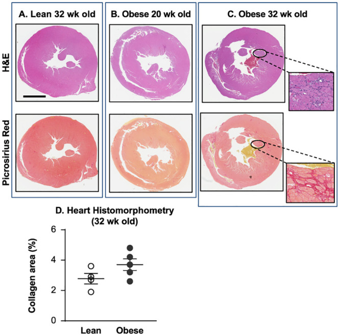
Fig 4. 20-week-old obese ZSF1 male rats did not exhibit histopathological events in heart tissue.

Histopathological evaluation of the hearts from lean (32-week-old, n = 4) and obese ZSF1 rats at the ages of 20 (n = 9) and 32 (n = 5) weeks demonstrated no evidence of fibrosis or chronic inflammation in the hearts from lean (A) or obese (B) 20-week-old ZSF1 males, but the hearts from 32-week-old obese rats (C) exhibited mild interstitial fibrosis with infiltration by mononuclear inflammatory cells in the myocardium; percentage of the myocardial area occupied by collagen (D) in 32-week-old obese males was increased, but did not achieve statistical significance vs the lean group. Fixed heart tissues were stained with standard hematoxylin and eosin or Picrosirius red; bar = 5 mm.
