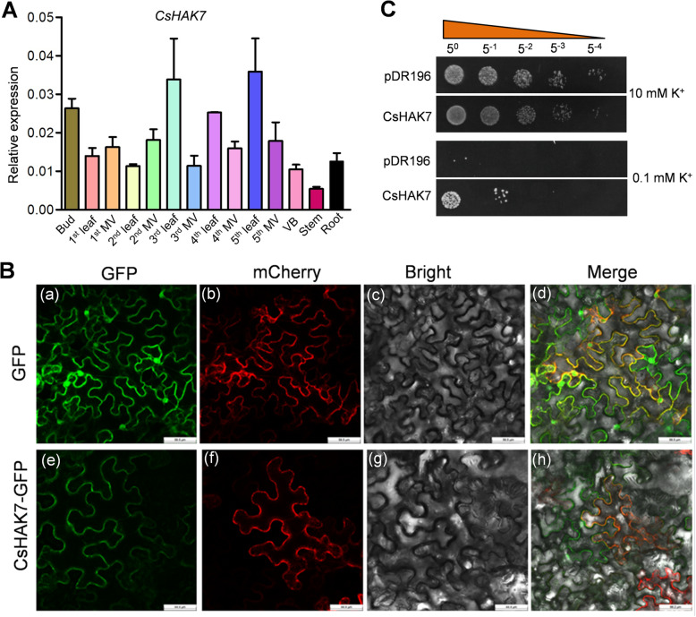
Fig. 9.
Tissue-specific expression pattern, subcellular localization and yeast complementation analysis of CsHAK7. a Expression pattern of CsHAK7 in indicated tissues of tea plant. MV, major vein; VB, vascular bundle, peeled from the stem. CsGAPDH was an internal control. Data are mean ± SE (n = 3). b Confocal laser scanning microscopy images showed tobacco leaf epidermal cells transiently expressing either GFP or CsHAK7::GFP together with AtPIP2A:mCherry (plasma membrane maker, Nelson et al., 2007). (a), (e), Confocal images via the GFP channel only. (b), (f), Confocal images of the red mCherry fluorescence marking the PM position. (c), (j), Bright field. (d), (h), Merged images of GFP (green) and mCherry RFP (red) together with bright filed. Scale bar, 45 μm. c Yeast complementation assay of K+ acquisition by the K+ uptake-defective yeast mutant (R5421) complemented with CsHAK7. Growth status of R5421 cells expressing CsHAK7, empty vector (pDR196), on AP solid medium containing 10 or 0.1 mM K+. The 1:5 serial dilutions of yeast cells were spotted on the AP solid medium and then incubated at 30 °C for 3–5 d

