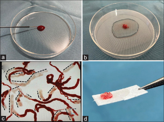
Figure 1.

The sample isolation processing by stereomicroscopy process for a EUS-fine-needle biopsy sample from a patient with gastrointestinal stromal tumor. (a) The sample in the puncture needle was initially pushed out onto a Petri dish by compressing the air in the syringe and then using a stylet. (b) The vermiform tissue component from the Petri dish was immersed in 10% neutral buffered formalin solution in another previously prepared Petri dish. (c) The stereomicroscopically visible white core lengths (black dashed lines) were measured using a scale on the stereomicroscope monitor screen. (d) The white and red samples were dissected using injection needles and closely aligned on separate filter papers. The photo is a white sample. SIPS: Sample isolation processing by stereomicroscope, EUS-FNB: EUS-guided fine-needle biopsy, GIST: Gastrointestinal stromal tumor, SVWC: Stereomicroscopic visible white core
