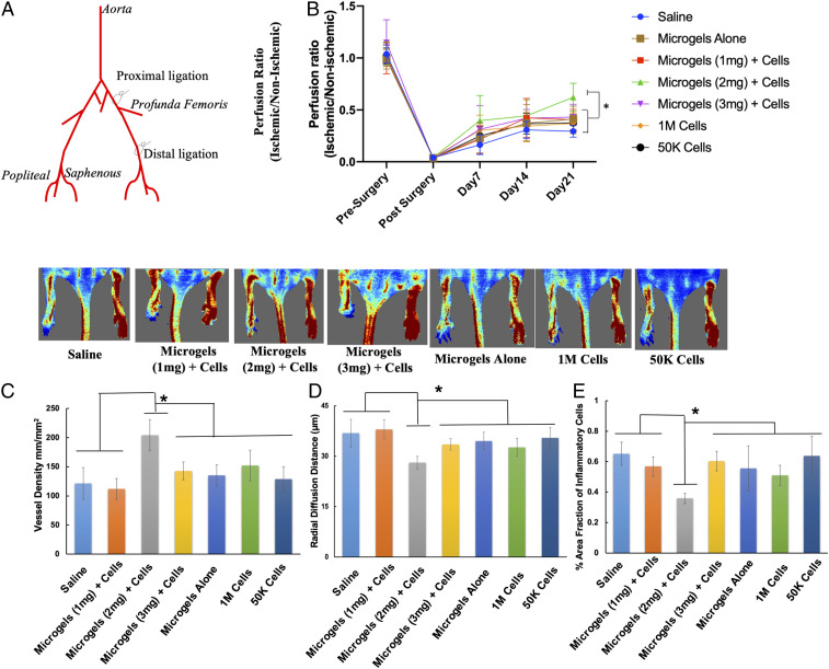
Fig. 6.
Improved blood flow in mice treated with hMSCs embedded in microgels at a low cell dose. (A) Schematic shows the double-ligation sites on the femoral artery in the hindlimb of a BALB/c nude mouse model. (B) Laser Doppler evaluation of the ischemic (left) and nonischemic (right) hindlimbs at day 21 (n = 12 per group, P < 0.05). (C and D) hMSC-embedded 2-mg⋅mL−1 microgels showed significantly higher capillary density (C) and a lower radial diffusion distance (D) compared to control groups at day 21. (E) Significantly lower infiltration of inflammatory cells was observed in 2-mg⋅mL−1 microgels with hMSCs compared to the rest of the treatment groups. (n = 12 per group, P < 0.05). *P < 0.05.

