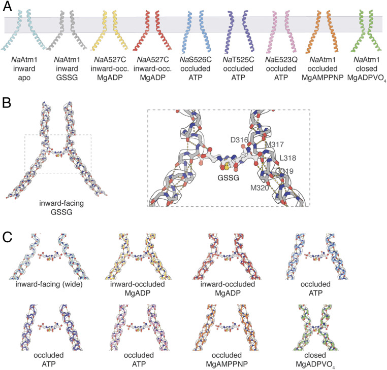
Fig. 4.
TM6 comparisons of NaAtm1. (A) TM6 (residues 300 to 340) conformations observed in different NaAtm1 structures. (B) Hydrogen-bonding interactions between GSSG and TM6 main chain groups from residues in the kink region of the primary binding site [residues 316 to 320 (PDB ID: 4MRS)]. (C) Potential interactions between GSSG and residues 316 to 320 in other NaAtm1 structures. The approximate location of GSSG is obtained from the structural alignments of TM6s in different structures to the TM6 of the inward-facing conformation of NaAtm1 (PDB ID: 4MRS). Hydrogen-bonding interactions are shown by yellow dashes and the GSSG positions are shown by ball and sticks in B and C. TM6s in the NaAtm1 wide-open inward-facing structure are shown in cyan, the NaAtm1 inward-facing structure (PDB ID: 4MRS) in gray, the NaA527C inward-occluded structure #1 in yellow, the NaA527C inward-occluded structure #2 in red, the NaS526C occluded structure in blue, the NaT525C occluded structure in purple, the NaE523Q occluded structure in pink, the NaAtm1 MgAMPPNP-bound occluded structure in orange, and the NaAtm1 MgADPVO4-bound closed structure in green.

