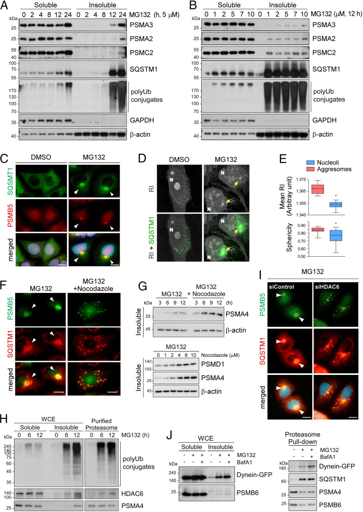
Fig. 1.
Treatment with proteasome inhibitors led to the accumulation of proteasomes in the insoluble aggresome via microtubule-based transport. (A) Accumulation of proteasome subunits in the insoluble fraction of mouse embryonic fibroblasts (MEFs) after treatment with 5 μM MG132 for the indicated periods (0 to 24 h). Whole cell extracts (WCEs) were separated into Triton X-100–soluble (soluble) and pellet fractions (insoluble) and subjected to SDS/PAGE/immunoblotting (IB). (B) As in A, except that the cells were treated with various concentrations of MG132 for 12 h. (C) Representative images of A549 cells treated with MG132 (10 μM, 12 h). Immunofluorescent staining (IFS) with anti-SQSTM1 (green) and anti-PSMB5 (red) antibodies. Nuclei were counterstained with DAPI (blue). (D) Refractive index (RI)-based holotomographic images of MG132 (5 μM, 12 h)-treated cells. Distinct juxtanuclear inclusion bodies from RI images are colocalized with SQSTM1-positive signals (green) from IFS. (E) Quantitative analysis of tomographic images of the aggresomes (mean RI and sphericity) compared with those of nucleoli from the identical cells. A box-and-whisker plot with n = 29. (F) Inhibition of aggresome formation in A549 cells by cotreatment with nocodazole (2 μM) and MG132 (5 μM) for 12 h. (G, Top) As in F, except that WCEs were fractionated into detergent-soluble and -insoluble fractions. (Bottom) A549 cells were treated with 5 μM along with indicated final concentrations of nocodazole for 12 h. Insoluble fractions were fractionated from WCEs and analyzed by SDS/PAGE/IB. (H) Direct interaction between HDAC6 and a proteasome was stronger with MG132 treatment. HEK293 cells stably expressing biotin-tagged CP subunit PSMB2 were treated with MG132 (5 μM) for 6 h or 12 h. Human proteasomes were affinity-purified from WCEs and then analyzed by SDS/PAGE/IB. (I) A549 cells were transfected with 20 nM siRNA for silencing HDAC6 (siHDAC6) or with scrambled (control) siRNA (siControl) for 48 h and then treated with 5 μM MG132 for 12 h. The cells were fixed and subjected to co-IFS with anti-PSMB5 (green) and anti-SQSTM1 (red) antibodies. (Scale bars: 10 μm.) (J) GFP-tagged dynein was transiently overexpressed in HEK293-PSMB2-biotin cells in the presence of 5 μM MG132 for 12 h and/or 100 nM bafilomycin A1 (BafA1) for 4 h. WCEs (Left) were subjected to affinity purification of proteasomes with streptavidin (Right), followed by IB with indicated antibodies.

