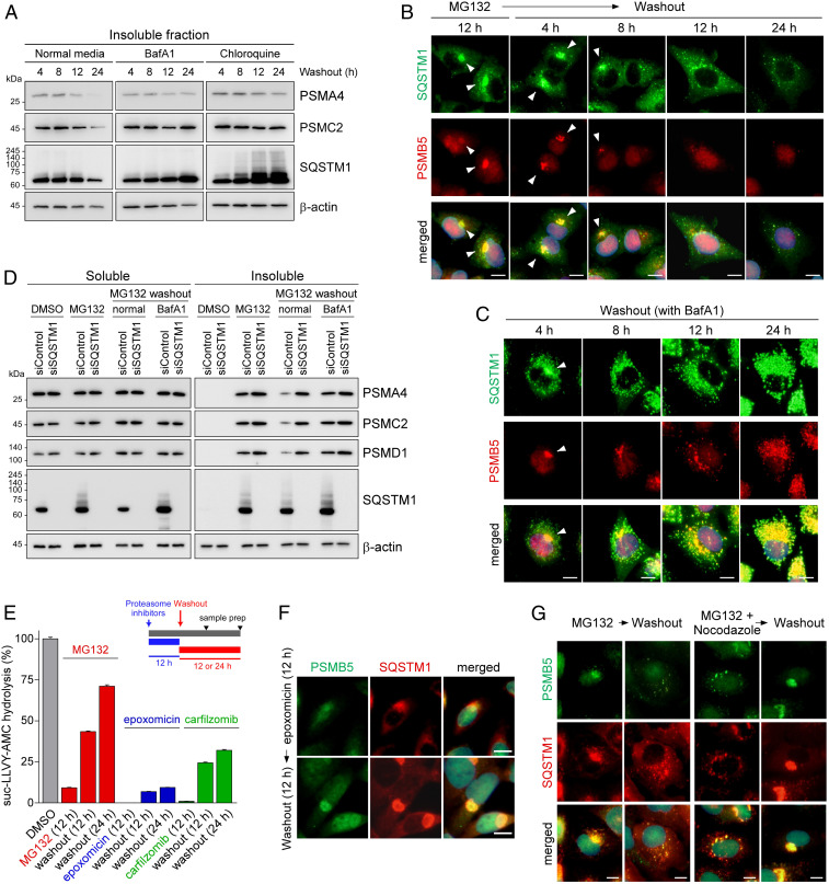
Fig. 2.
Inhibited proteasomes accumulated in the aggresome are cleared via the autophagy–lysosome system. (A) Washing out MG132 with either a normal medium or medium containing autophagy inhibitors, such as BafA1 (100 nM) and chloroquine (25 μM), for 4, 8, 12, or 24 h. HEK293 cells were treated with MG132 (5 μM) for 12 h to induce aggresome formation before the washout experiment. Immunoblotting analysis of a CP subunit (PSMA4), an RP subunit (PSMC2), SQSTM1, and β-actin was conducted after the isolation of the detergent-insoluble fraction of WCEs. (B and C) As in A, except that IFS analysis was performed with anti-PSMB5 (red) and anti-SQSTM1 (green) antibodies in the absence (B) or presence (C) of BafA1. (D) A549 cells were transfected with either siControl or siSQSTM1. After 24 h, cells were treated with 5 μM MG132 for 12 h and then washed out with either a normal medium or a medium containing BafA1 for additional 18 h. A detergent-soluble and -insoluble fraction was isolated and subject to SDS/PAGE/IB analysis. (E) The proteasome activity was measured using hydrolysis of fluorogenic suc-LLVY-AMC after treatment with 5 μM MG132, 0.5 μM epoxomicin, or 0.1 μM carfilzomib for 12 h (mean ± SD from three independent experiments). (F) Contrary to the results from MG132-treated samples, proteasome-positive aggresomes after treatment with irreversible proteasome inhibitor epoxomicin (100 nM, 12 h) did not dissipate even after 12 h inhibitor washout. (G) IFS images of A549 cells after 12 h washout following 12 h MG132 treatment (5 μM) or MG132 plus nocodazole (2 μM) cotreatments. (Scale bars: 10 μm.)

