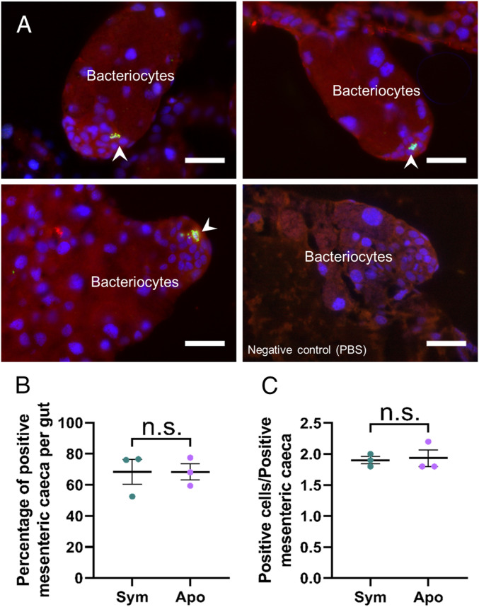
Fig. 4.
Putative epithelial stem cells are present at the apices of forming bacteriomes. (A) PH3 localization by immunostaining in forming bacteriomes in pupae. A negative control was included (Right, PBS). PH3 is specifically localized in clusters of small cells at the apices of forming bacteriomes (arrowheads), indicating that they might be stem cells. Red: autofluorescence; green: PH3; blue: DAPI. (Scale bars, 25 µm.) (B) Percentages of bacteriome containing PH3+ cells in whole guts from symbiotic and aposymbiotic pupae. The means and SEs for three independent replicates are represented. No significant difference was found between symbiotic and aposymbiotic individuals, based on a Welch t test. (C) Numbers of PH3+ cells per positive bacteriome in symbiotic and aposymbiotic pupae. The means and SEs for three independent replicates are represented. No significant difference was found between symbiotic and symbiotic individuals, based on a Welch t test; n.s., not significant.

