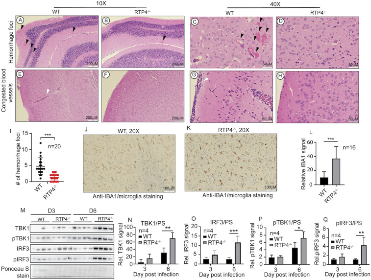
Fig. 8.
Brain pathological changes, microglia activation, and IFN-I response in mice infected with P. berghei ANKA. (A–D) Images showing hemorrhage foci at the cerebellum. (E–H) Blood vessels with iRBCs and host white cells from the cerebellum. (I) Numbers of hemorrhage foci from 20 randomly selected microscopic images from the cerebellum of P. berghei-infected WT and Rtp4−/− mice. Brain samples from mice at day 6 p.i. were fixed in 10% buffered formalin, embedded in paraffin, and sequential coronal sections of the whole brain were stained with hematoxylin and eosin (H&E) or antibodies as described. H&E-stained brain tissue sections were examined under a light microscope at 10× and 40× magnifications. (J and K) Anti-IBA1 staining (brown color) of microglia in the brain from cerebellar cortex of infected WT (J) and Rtp4−/− (K) mice, as described in Materials and Methods. (L) Quantitative microglia signals as shown in J and K were scanned from anti-IAB1–stained images (16 each) of brain tissues of WT and Rtp4−/− mice at day 6 p.i. with P. berghei using ImageJ. (M) Western blot of protein expression and phosphorylation of TBK1 and IRF3 in microglia purified from the whole brains of infected WT and Rtp4−/− mice (n = 4) at days 3 and 6 p.i. with P. berghei ANKA. Scanned signals of Ponceau S-stained total proteins as loading controls. (N–Q) Plots of scanned protein and phosphorylation signals of TBK1 and IRF3 from M. For I, L, and N–Q, two-way ANOVA, mean and SD: *P < 0.05; **P < 0.01; ***P < 0.001.

