Fig. 7.
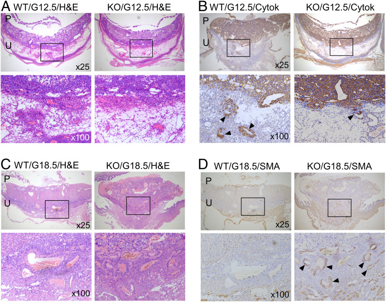
Trophoblast invasion and spiral artery remodeling in Klf17−/− mice. (A and B) Placental (P) and uterine (U) sections from WT and Klf17−/− (KO) mice at G12.5 were stained with H&E (A) or immune-stained with an anti-cytokeratin 18 (Cytok) antibody (B). Boxed areas in low-magnification (×25) images (Top row) are shown at a higher magnification (×100) (Bottom row). Endovascular trophoblast invasion in decidual sections are indicated by arrowheads. (C and D) Placental (P) and uterine (U) sections from WT and KO mice at G18.5 were stained with H&E (C) or immune-stained with an anti-SMA antibody (D). Boxed areas of low-magnification (×25) images (Top row) are shown at a higher magnification (×100) (Bottom row). Positive SMA staining in decidual arteries in KO mice are indicated by arrowheads. Data are representative of studies in at least five mice per group.

