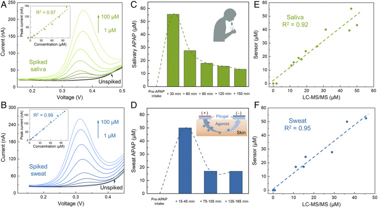
Fig. 3.
Nafion/H-BDDE-enabled ex situ APAP quantification in noninvasively retrieved biofluid samples of (A, C, and E) saliva and (B, D, and F) sweat. (A and B) Differential pulse voltammograms of unspiked and spiked (with 1, 5, 10, 20, 40, 60, 80, and 100 μM APAP) saliva (A) and sweat (B) samples. (Insets) The corresponding analytical framework-extracted peak current. (C and D) Sensor-measured APAP concentration in the saliva (C) and sweat (D) samples of a human subject, collected before and at intermittent time points after the oral administration of a medication containing 650 mg APAP. (Insets) The schematics of saliva collection and iontophoresis-based sweat stimulation. (E and F) Sensor-measured APAP concentrations in saliva (E) and sweat (F) samples versus the corresponding LC-MS/MS readouts.

