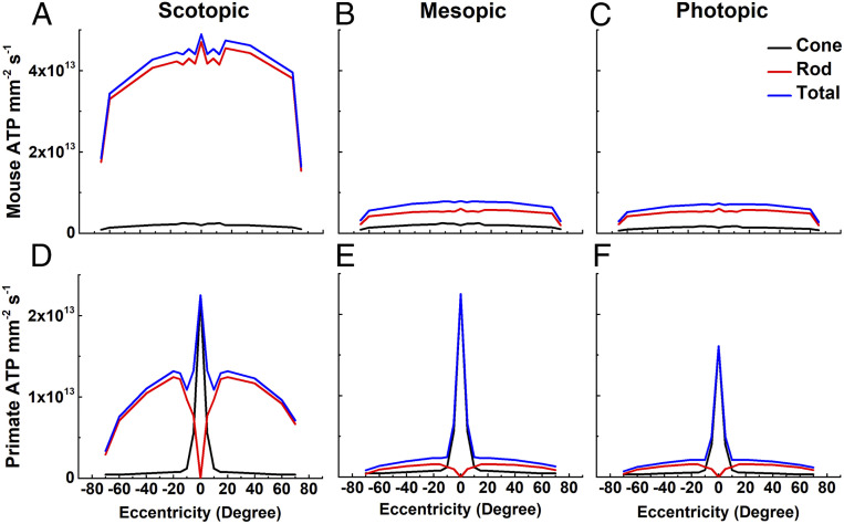
Fig. 3.
ATP consumption across retinal eccentricities. We used total ATP consumption for rods and cones from Fig. 2 together with measurements of the density of photoreceptors as a function of retinal eccentricity (mouse, 32; human, 36) to calculate the metabolic demand of the outer retina in mouse (A–C) and human (D–F) in three ambient light intensities: scotopic (darkness; 0 effective ϕ cell−1s−1) (A and D); mesopic (12,600 effective ϕ rod−1s−1 or 12,500 effective ϕ cone−1s−1) (B and E); and photopic (8.8 × 10⁷ effective ϕ rod−1s−1 or 8.7 × 10⁷ effective ϕ cone−1s−1) (C and F). The same values were used for cone currents, even though foveal cone outer segments are three times longer than mouse cones (38, 39) and likely have larger circulating currents. No attempt was made to take the difference in the number of synaptic ribbons in the pedicles between foveal and peripheral cones into consideration (40); these factors would only increase the difference in ATP consumption between rods and cones.

