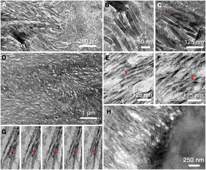
Fig. 2.
TEM characterization of KLK4−/− mice incisor sections before and after demineralization. (A) TEM image near the DEJ region before demineralization. (B) Close-up image shows individual apatite crystals. (C) Some apatite crystals present with a typical flexible ribbon-like morphology (indicated with red arrow). (D) TEM image of demineralized enamel region reveals an extensive protein matrix network. (E) Close-up images reveal that the protein matrix network is composed of individual protein nanoribbon structures. Some of these share the unique central dark line (indicated with red arrow) as observed with rH146 nanoribbons in Fig. 1G. (F) Twisting of the nanoribbon structures (indicated with red arrow) is frequently observed. (G) From left to right, a series of TEM images of the same protein nanoribbon (indicated with red arrow) with increasing tilting angles counterclockwise shows the transition from the ribbon top view to its side view. (H) TEM image near the DEJ region after demineralization. Collagen structures can be observed in the demineralized dentin region, and amelogenin nanoribbons can be seen to grow out from the dentin surface directly.

