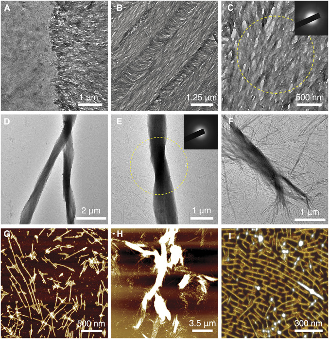
Fig. 3.
PILP-induced mineral formation on both tissue-based amelogenin nanoribbons and rH146 amelogenin nanoribbons. (A) TEM image near the DEJ region after incubating demineralized KLK4−/− mouse incisor section with PILP solution at 37 °C for 1 h. (B) TEM image of the demineralized KLK4−/− mouse incisor section after incubating in PILP solution for 4.5 h. (C) Close-up TEM image of amorphous mineral coating over amelogenin ribbons; yellow circle corresponds to area of SAED analysis (Inset), which indicates absence of crystalline phase. (D) TEM image of the rH146 nanoribbon and PILP-solution mixing sample after incubating for 15 d shows the formation of large bundled structures. (E) TEM image of mineralized rH146 ribbon bundle; yellow circle corresponds to area of SAED analysis, SAED (Inset) shows that mineral within the bundled structures remained amorphous at 15 d. (F) Close-up image reveals the process where individual nanoribbons associate together and align to form larger bundles. (G) AFM image of the rH146 nanoribbon and PILP-solution mixing sample. PILP droplets can be seen to bind with rH146 nanoribbons. (H) AFM image of the rH146 nanoribbon and PILP-solution mixing sample shows the formation of large bundles. (I) Three-dimensional rending of AFM image reveals formation of bridges between nanoribbons when exposed to PILP solution, which generates a unique pattern on the mica substrate.

