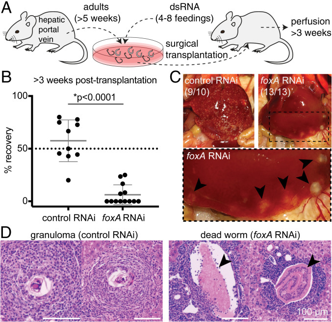
Fig. 4.
The esophageal gland is required for parasite survival inside the mammalian host. (A) Experimental scheme for the transplantation of adult schistosomes after RNAi. (B) Worm recovery >3 wk after the surgical transplantation. Each dot represents one host mouse. Mean ± SD. Statistical analysis: Welch’s t test. Raw numbers are shown in SI Appendix, Table S1. (C) Representative images of whole livers harvested from mice transplanted with control (Left) or foxA RNAi (Right) parasites after >3 wk. Magnified region inside the dotted box is shown below. Black arrowheads: dead worms flushed into the liver. The numbers indicate how many livers show the respective phenotype. (D, Left) Representative H&E staining of granulomas in liver sections of mouse transplanted with control RNAi parasites. (Right) Representative H&E staining showing a cross section of dead worms (arrowheads) in liver sections of mouse transplanted with foxA RNAi parasites. See SI Appendix, Fig. S6B for additional H&E staining.

