Fig. 6.
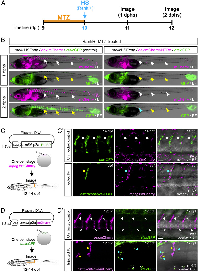
Osteoblast-derived Cxcl9l is sufficient for macrophage recruitment and osteoclast differentiation in the absence of Rankl induction. (A) Experimental timeline for osteoblast ablation. osx:mCherry-NTRo larvae were treated with Metronidazole (MTZ) at 9 dpf for 24 h, and heat shock was applied at 10 dpf to induce Rankl expression. (B) Osteoblasts (magenta) and osteoclasts (green) were imaged at 1 and 2 dphs. osx:mCherry larvae treated with Mtz were used as controls. Osteoblasts and osteoclasts form normally in controls (white and yellow arrows, respectively) but are absent in osteoblast-ablated larvae (white and yellow arrowheads; blue arrowhead highlights remaining osteoclasts). (C) Strategy for macrophage analysis after ectopic cxcl9l expression in osx osteoblasts. mpeg1:mCherry transgenic embryos were injected with osx:cxcl9l-p2a-EGFP plasmid and I-SceI meganuclease at one-cell stage, raised to 12 to 14 dpf, and screened for mosaic osx:cxcl9l-p2a-EGFP expression in osteoblasts located at vertebral bodies. (C') Confocal images showing single macrophages present at neural arches of noninjected osx:GFP/mpeg1:mCherry control larvae (Top; white arrows, n = 5 fish). Enhanced recruitment of macrophages (cyan arrows) toward cxcl9l-p2a-EGFP expressing cells (yellow arrow) in neural arch of osx:cxcl9-p2a-EGFP injected larva (Bottom; n = 7/7 fish). (D) Strategy for osteoclast analysis upon ectopic cxcl9l expression in osx-expressing osteoblasts. ctsk:GFP transgenic embryos were injected with osx:cxcl9l-p2a-mCherry plasmid and I-SceI meganuclease at one-cell stage, raised to 12 to 14 dpf, and screened for mosaic osx:cxcl9l-p2a-mCherry expression in osteoblasts at vertebral bodies. (D') Confocal images showing absence of osteoclast formation (white arrowheads) in the vertebral bodies of uninjected osx:mCherry/ctsk:GFP larvae (Top; control, n = 7 fish). Ectopic formation of ctsk:GFP-expressing osteoclasts (cyan arrowheads) in vertebral bodies upon mosaic cxcl9l-p2a-mCherry expression in osx cells (yellow arrowheads; Bottom; n = 6/6 fish; 4/6 fish showed ectopic osteoclast formation both close to and distant from Cxcl9l-expressing cells, and 2/6 fish showed ectopic osteoclast formation at a distance from Cxcl9l-expressing cells). Asterisks indicate autofluorescent pigment cells at yolk region. (Scale bars, 50 μm.)

