Fig. 1.
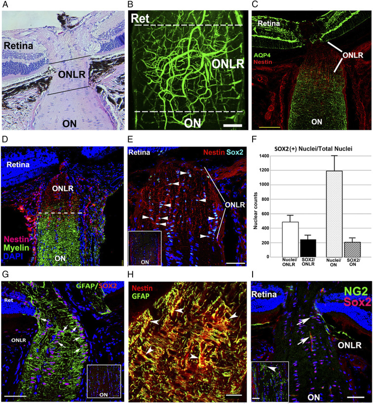
Rodent ONLR characterization. (A) H&E longitudinal section, mouse ONLR. (B) Two-photon micrograph of fluorescein-filled rodent (rat) ONLR vessels. ONLR vasculature forms a plexus with contributions from retina (Ret) and ON. (C) AQP4 expression in the retina, ONLR, and ON. AQP4 (in green)/Nestin (in red). There is reduced ONLR-AQP4, compared with retina or distal ON. (D) Nestin localization in the ONLR. Nestin (red)/myelin (O4 antibody; green) localization define the ONLR. Nestin expression declines anterograde, with early oligodendrocyte development only below the ONLR. (E) Increased SOX2 expression in the ONLR. SOX2(+) nuclei are qualitatively increased in the nestin-enriched ONLR (arrowheads), and present in reduced numbers in the distal ON (Fig. 1 G, Inset: ON). (F) SOX2 nuclear quantification in ONLR and ON. ONLR is enriched in SOX2 nuclei, in terms of both area (Left bars), and SOX2:total nuclei (Right bars). (G) ONLR-SOX2/GFAP expression. GFAP surrounds SOX2(+) nuclei, suggesting that ONLR SOX2(+) cells express both proteins. Distal ON (Inset) reveals that cells with SOX2(+) nuclei lack GFAP expression. (H) Nestin and GFAP colocalize in the ONLR. Red: Nestin; green: GFAP. (Scale bar: 20 μm.) (I) NG2(+)/Sox2(+) OPCs are present only distal to the mouse ONLR. NG2(+) OPCs (green) are only seen in the anterior ON below the ONLR. NG2(+) capillaries/(arrow) are present in the ONLR. Inset: NG2(+) cells have a Sox2(+) nucleus (in red). (Scale bar in H: 20 μm; C, E, and G: 50 μm; I: 100 μm; and Inset in I: 50 μm.)

