Fig. 2.
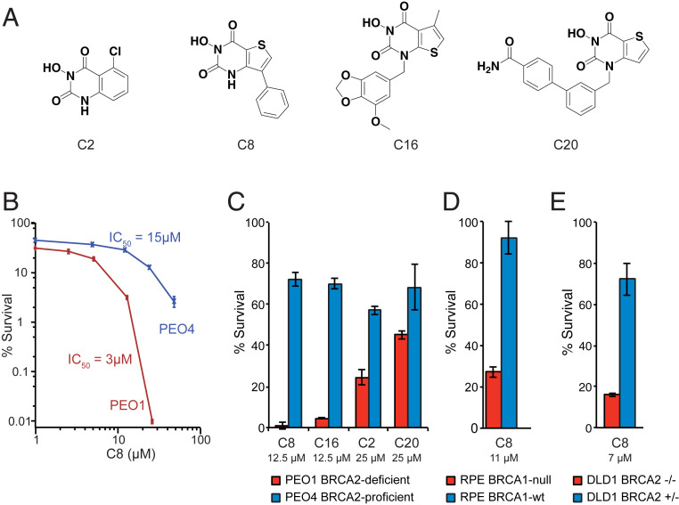
BRCA-deficient cancer cell lines are sensitive to FEN1 inhibition. (A) Structures of the synthesized FEN1 inhibitors. (B) Clonogenic survival of BRCA2-deficient PEO1 and BRCA2-revertant PEO4 cells demonstrates that PEO1 cells are more sensitive to a 3-d exposure to the C8 FEN1 inhibitor. (C) Clonogenic survival demonstrates that PEO1 cells are more sensitive than PEO4 cells to a 3-d exposure to all of the FEN1 inhibitors, with C8 and C16 causing the greatest reduction in percent survival. C8 and C16 were tested at 12.5 μM and C2 and C20 were tested at 25 μM. (D) RPE1-hTERT p53−/− BRCA1-KO cells are more sensitive to a 3-d exposure to 11 μM C8 than the parental BRCA1–wild-type (wt) cells. (E) DLD1 BRCA2−/− cells are more sensitive to a 3-d exposure to 7 μM C8 than the parental BRCA2-proficient cells. Error bars represent the standard deviation.

