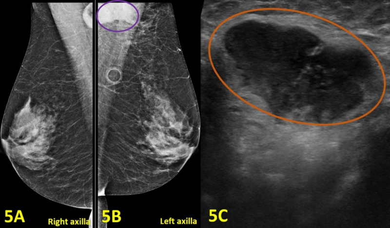
Figure 5. Imaging of Bilateral Breasts, Bilateral Axillae, and Left Axillary Lymphadenopathy.
(A) Mammogram of the right breast and axilla shows no abnormal findings. (B) Diagnostic mammogram of the left breast and axilla shows an enlarged lymph node with adjacent abscess measured 30 x 17 mm (purple oval). (C) Left axilla diagnostic ultrasound demonstrates an enlarged axillary lymph node with cortical thickening measuring 30 mm (orange oval).

