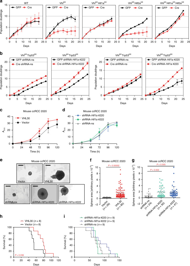
Fig. 2. HIF-1α is dispensable for cellular proliferation and for allograft tumour formation.
a 3T3 proliferation assays of MEFs derived from mice of the indicated genotypes infected with adenoviruses expressing GFP or Cre. Mean ± std. dev. are derived from three independent cultures. b 3T3 proliferation assays of MEFs derived from Vhlfl/flTrp53fl/fl mice infected with non-silencing shRNA (shRNA-ns) or shRNA against Hif1a (shRNA-Hif1a #1 and shRNA-Hif1a #2), followed by infection with adenoviruses expressing GFP or Cre. Mean ± std. dev. are derived from three independent cultures. c, d Proliferation assays of mouse ccRCC cell line 2020 expressing empty vector control or human pVHL30 c or non-silencing shRNA (shRNA-ns) or shRNA against Hif1a (shRNA-Hif1a #1 and shRNA-Hif1a #2) d. Mean ± std. dev. are derived from two independent experiments each with replicates of six cultures. e–g Representative images (scale bars depict 200 μm) e and size distributions f, g of spheres formed by the cells described in c, d when grown in non-adherent cell culture plates. Mean ± std. dev. of the total number of colonies pooled from three independent experiments are shown, P values were calculated by two-sided Student’s t test. h, i Survival of mice following subcutaneous allograft tumour assays of the cells described in c into SCID-Beige mice. P values were calculated by two-sided log-rank Mantel–Cox test.

