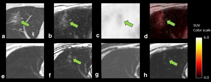
Fig. 6.
A case showing lung adenocarcinoma in a 61-year-old man after surgery for pharyngeal cancer. CT (a) showed a 7.3-mm-sized lesion with ground-glass opacity component. ZTE (b) can reveal these CT characteristics, leading to correct diagnosis of lung cancer by the combined findings of the FDG uptake on PET (SUVmax 3.9, c) and fused images (d). Dixon sequence (e, in phase; f, out of phase; g, fat; and h, water image, respectively), however, failed to depict the characteristics of ground-glass opacity, resulting in the diagnosis of differentiation as equivocal

