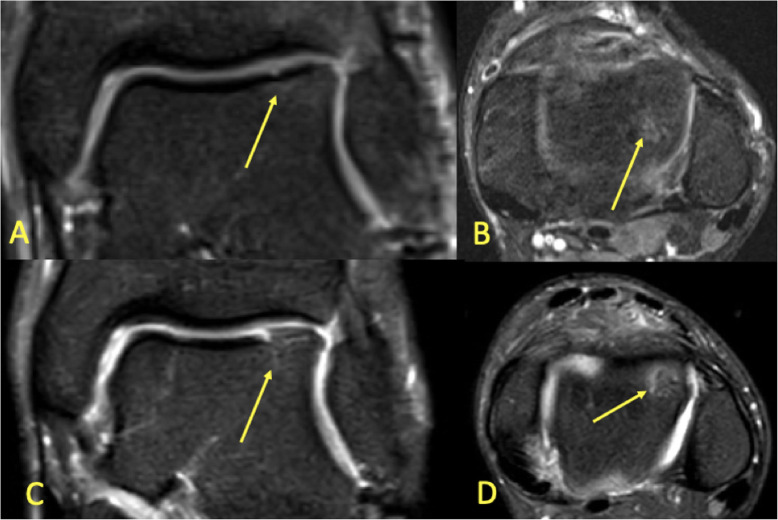
Fig. 17.
A 19-year-old handball player with 3-month history of ankle pain imaged for suspected anterior tibiofibular ligament rupture and stress fracture. a, b On the initial MRI, the cartilage lesion (arrow) was missed, and neither an anterior tibiofibular ligament rupture nor a stress fracture was detected. c, d Because of persistent pain preventing training, MRI was repeated after 8 months. A chondral lesion with subchondral BME was visible and could be identified retrospectively on the previous MRI (arrows)

