Figure 3.
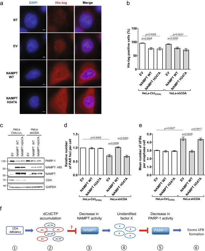
The low levels of PARP-1 activity in CDA-deficient cells are rescued by the overexpression of wild-type NAMPT (a) Representative immunofluorescence deconvoluted z-projection images showing DAPI and His-tag staining in HeLa-Ctrl(CDA) cells not transfected (NT) or transiently transfected with pPM-C-His empty vector (EV), or with a pPM-C-His construct expressing wild-type NAMPT (NAMPT WT) or mutated NAMPT (NAMPT H247A). Nuclei were visualized by DAPI staining (blue) and the His-tag was visualized with Alexa Fluor 555 (red). Scale bar: 5 µm. (b) Percentage of His-tag-positive cells among HeLa-Ctrl(CDA) and HeLa-shCDA cells transiently transfected with EV, NAMPT WT or NAMPT H247A. The data shown are means ± SD from four independent experiments. (c) PARP-1, NAMPT, NAMPT-HIS and CDA proteins levels assessed by immunoblotting in HeLa-Ctrl(CDA) and HeLa-shCDA cells transiently transfected with EV, NAMPT WT or NAMPT H247A. (d) Relative number of PAR foci in HeLa-Ctrl(CDA) and HeLa-shCDA) cells transiently transfected with EV, NAMPT WT or NAMPT H247A. The data shown are means ± SD from four independent experiments (> 800 cells per condition). (e) Mean number of UFBs per anaphase cell, for HeLa-Ctrl(CDA) and HeLa-shCDA cell lines transiently transfected with EV, NAMPT WT or NAMPT H247A. Error bars represent means ± SD from three independent experiments (> 120 anaphase cells per condition). The significance of differences was assessed in Student’s t-tests. (f) (1) CDA deficiency leads to (2) intracellular dC/dCTP accumulation that (3) decreases NAMPT activity, directly or indirectly, leading to the (4) intracellular accumulation of an as yet unidentified factor X (5) lowering basal PARP-1 activity, causing (6) excess UFB formation.

