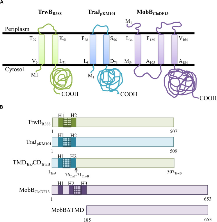
FIGURE 1.
(A) Predicted membrane topology of TrwBR388, TraJpKM101, and MobBCloDF13 proteins. Membrane topology of the different T4CPs was predicted using Topcons software. The black lines represent the inner bacterial membrane. M1, amino-terminus; COOH, carboxy-terminus. The first and last residues of each transmembrane helix are shown indicating their position in the sequence. Proteins from R388, pKM101, and CloDF13 plasmids are shown in green, blue, and purple, respectively. (B) Schematic representation of the different T4CPs and their variants used in the present study. Proteins from R388, pKM101, and CloDF13 plasmids are shown in green, blue and purple, respectively. The transmenbrane α-helices (H) and the small periplasmic loops connecting α-helices are indicated in dark boxes and stripped boxes, respectively.

